INLAND HEART DOCTORS
  • Home
  • Services
    • Interventional Cardiology >
      • Watchman Device
      • MitraClip
    • Venous Reflux Disease
  • Providers
  • Patient Resources
    • New Patient Registration
    • Make a Payment
    • Billing Questions
    • Patient Portal
    • Request Records
    • Insurance
    • Patient Education
  • Locations

​Interventional Cardiology

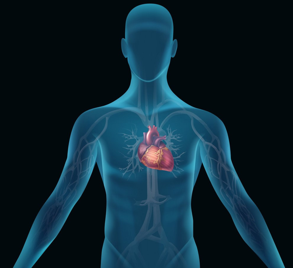
​Our Board-Certified Cardiovascular experts provide each patient with a full spectrum of cardiac treatments available. Our team not only focuses on addressing challenges, but also in setting goals for therapy, helping to achieve Optimal Cardiovascular Health.
​Whether evaluation, diagnosis or management of Cardiovascular Diseases, we work closely with patients, their families and primary care providers to build an individualized treatment plan according to each unique need. We are honored to have the opportunity of being your trusted partner in complete Cardiovascular health
Picture

​Cardiovascular Procedures

While prevention and minimal invasive treatments are our goal, some Interventions are necessary to address some Cardiovascular Conditions.  Here are some common procedures as a point of reference to help you understand their purpose in your overall treatment plan:
Cardiac Catheterization Coronary Intervention
What is a Cardiac Catheterization? 
Cardiac Catheterization, also called Cardiac Cath or Coronary Angiogram, is an invasive imaging procedure that allows your doctor to evaluate your heart function. 

Cardiac Catheterization is used to evaluate or confirm the presence of Coronary Artery Disease, Valve Disease, or disease of the Aorta. It can be used to evaluate heart muscle function and to determine the need for further treatment such as an interventional procedure or Coronary Artery Bypass Graft, or CABG, surgery. 
During a Cardiac Catheterization, a long, narrow tube called a catheter is inserted through a plastic introducer sheath (a short, hollow tube that is inserted into a blood vessel in your arm or leg). The catheter is guided through the blood vessel to the Coronary Arteries with the aid of a special x-ray machine.  

Contrast material is injected through the catheter and x-ray movies are created as the contrast material moves through the heart’s chambers, valves and major vessels. This part of the procedure is called a Coronary Angiogram (or Coronary Angiography). The digital photographs of the contrast material are used to identify the site of the narrowing or blockage in the Coronary Artery. Additional imaging procedures, called Intra-Vascular Ultrasound (IVUS) and fractional flow reserve (FFR), may be performed along with Cardiac Catheterization in some cases to obtain detailed images of the walls of the blood vessels. 

With IVUS, a miniature sound-probe (transducer) is positioned on the tip of a Coronary Catheter. The catheter is threaded through the Coronary Arteries and, using high-frequency sound waves, produces detailed images of the inside walls of the arteries. IVUS produces an accurate picture of the location and extent of plaque.
​Peripheral Angiogram /Angioplasty / Intervention
What is an Interventional Procedure?
An Interventional Procedure is a non-surgical treatment used to open narrowed Coronary Arteries to improve blood flow to the heart. An Interventional Procedure can be performed during a diagnostic Cardiac Catheterization when a blockage is identified, or it may be scheduled after a Catheterization has confirmed the presence of Coronary Artery Disease.
An Interventional Procedure starts out the same way as a Cardiac Catheterization. Once the catheter is in place, one of these interventional procedures is performed to open the artery: Balloon Angioplasty, Stent Placement, Rotablation or Cutting Balloon.
  • Balloon Angioplasty: A procedure in which a small balloon at the tip of the catheter is inserted near the blocked or narrowed area of the Coronary Artery. The technical name for Balloon Angioplasty is Percutaneous Transluminal Coronary Angioplasty (PTCA) or Percutaneous Coronary Intervention (PCI). When the balloon is inflated, the fatty plaque or blockage is compressed against the artery walls and the diameter of the blood vessel is widened (dilated) to increase blood flow to the heart. 
  • Balloon Angioplasty with Stenting: In most cases, Balloon Angioplasty is performed in combination with the stenting procedure. A stent is a small, metal mesh tube that acts as a scaffold to provide support inside the Coronary Artery. A balloon catheter, placed over a guide wire, is used to insert the stent into the narrowed artery. Once in place, the balloon is inflated and the stent expands to the size of the artery and holds it open. The balloon is deflated and removed, and the stent stays in place permanently. During a period of several weeks, the artery heals around the stent. 

Why is a Cardiac Catherization done? 
Cardiac Catheterization is done to see if you have a heart problem. It can also be done as part of a procedure to correct a known heart problem.

If you're having Cardiac Catheterization as a test for Heart Disease, your doctor can:
• Locate narrowing or blockages in your blood vessels that could cause chest pain (Angiogram)
• Measure pressure and oxygen levels in different parts of your heart (Hemodynamic Assessment)
• Check the pumping function of your heart (Right or Left Ventriculogram)
• Take a sample of tissue from your heart (Biopsy)
• Diagnose heart defects present from birth (Congenital Heart Defects)
• Look for problems with your heart valves

Cardiac catheterization is also used as part of some procedures to treat heart disease.
These procedures include
:
• Widening a narrowed artery (Angioplasty) with or without stent placement
• Closing holes in the heart and fixing other congenital defects
• Repairing or replacing heart valves
• Opening narrow heart valves (Balloon Valvuloplasty)
• Treating irregular heart rhythms with ablation
• Closing off part of your heart to prevent blood clots
​Transcatheter Aortic Valve Replacement  (TAVR)
What is Aortic Stenosis?
Aortic Stenosis is a condition in which the Aortic Valve, the main valve connecting the heart to the rest of the body, does not open fully due to thickening of the valve leaflets. Valve disease can occur for several reasons, but age-related factors are the most common in the United States.
This leads to significantly increased load and work on the heart muscle and reduced blood flow to the rest of the body. Over time this can lead to symptoms of increased fatigue, decreased activity tolerance, chest discomfort, difficulty breathing, lightheadedness, dizziness and fainting.
A multidisciplinary team can provide a comprehensive evaluation to determine the optimal timing and treatment options for each patient depending on aortic stenosis severity and symptoms.
Once treatment is indicated, severe aortic stenosis can be treated with either minimally invasive (transcatheter) valve replacement or open surgical valve replacement.

What is the TAVR Procedure?
Transcatheter Aortic Valve Replacement, or TAVR, is a minimally invasive, catheter-based procedure to replace the diseased aortic valve in patients with severe aortic stenosis. Previously, the only treatment for aortic stenosis was open-heart surgical aortic valve replacement surgery (SAVR).
Over the past years, TAVR has been shown to be as effective as surgical aortic valve replacement in the treatment of severe aortic stenosis most patients and has rapidly become the treatment of choice for many patients.

Performed by a team led by an Interventional Cardiologist and a cardiac surgeon, TAVR uses a catheter to deliver a new prosthetic valve to the heart.
The valve can be delivered via several approaches depending on the team’s recommendation. The most common approach involves access via a blood vessel in the patient's groin (femoral artery). Once the new valve is perfectly positioned, it is opened, pushing aside the flaps of the original defective valve and effectively replacing it.
This procedure is also effective in the treatment of previously replaced failing tissue (bioprosthetic) surgical valves.

Who is a Good Candidate for TAVR?
Our Heart Team provides an extensive evaluation which culminates in a multidisciplinary discussion to determine individualized treatment options and can make recommendations with regards to the best treatment options.
Recent studies have shown benefit across the spectrum of surgical risk (low, intermediate, high and prohibitive risk for open heart surgery). As such, TAVR is now approved and shown to highly benefit patients regardless of their risk open heart surgery. The decision on the best treatment option now is based on individual patient factors and shared-decision making between the team and patient. While open heart surgery cannot always be avoided, we anticipate that TAVR will become the standard of care for all aortic stenosis patients in the near future.
Like all procedures there are certain risks associated with transcatheter aortic valve replacement.

Our Interventional Team can discuss with you the potential risks and benefits and help decide if this procedure is right for you.
​
To schedule an appointment to discuss Aortic Stenosis or other Heart Valve condition. Please call us at (951) 734-4880 for an appointment. 
​MITRACLIP® FOR DEGENERATIVE MITRAL REGURGITATION (DMR)
What is Mitral Valve Regurgitation?
Mitral Valve Regurgitation is a condition in which the heart’s mitral valve does not close tightly. As a result, blood flows backwards into the upper chamber (left atrium) from the lower chamber (left ventricle) as the heart contracts. This reduces the amount of blood that flows through your heart and to the rest of the body. Because of this backflow of blood, the heart has to work harder to pump blood from the left ventricle to the aorta, resulting in an enlarged left ventricle.

Help for Patients at High Risk for Surgical Mitral Valve Repair or Replacement
At INLAND HEART DOCTORS, our Interventional Team will provide the highest level of care for heart valve disease including leading-edge therapies like the MitraClip® If you have been told that you are at high risk for heart valve surgery, these minimally invasive procedures may be a good alternative.
Unlike traditional surgical interventions, the MitraClip® procedure does not require opening the chest and temporarily stopping the heart. 

Our Interventional Team can discuss with you the potential risks and benefits and help decide if this procedure is right for you.
​
To schedule an appointment to discuss Mitral Valve Regurgitation or other Heart condition.
Please call us at 
(951) 734-4880 for an appointment.  
VISIT OUT MITRACLIP PAGE TO FIND OUT MORE INFORMATION
Patent Foramen Ovale Repair (PFO)
​What is a Patent Foramen Ovale (PFO)? 
Before birth, a baby’s heart will have a hole with flap-like covering between the upper two chambers of the heart. This opening (Foramen Ovale) allows blood rich in oxygen from the mother to bypass the baby’s lungs which do not function until the baby is born. After birth, the flap-like covering will typically close the hole permanently. 
However, in about one out of every four individuals, the hole will remain open. This is called a Patent Foramen Ovale or PFO. From time to time, a PFO may permit blood to pass from the right side of the heart to the left side of the heart bypassing the normal route of going through the lungs first. Symptoms In most people, a PFO creates no symptoms and requires no treatment. However, in a small minority, a PFO may permit blood clots to pass from the right side of the heart to the left side possibly leading to a stroke.

What is a stroke? 
A stroke occurs when either the cells in the brain do not receive the oxygen needed to survive. When this happens, brain cells begin to die. The parts of the body controlled by the area of the brain damaged by the stroke may not function correctly. For instance, stroke can lead to problems speaking or moving.

What causes a Stroke? 
There are two main types of stroke. One type, called hemorrhagic stroke, occurs when injured blood vessels bleed into the brain tissue. This type of stroke happens most often in patients with high blood pressure. 

A second type of stroke is called ischemic stroke. This occurs when a blood vessel carrying blood to the brain is blocked. 

Many times, these strokes have an identifiable cause. One common cause is the buildup of plaque (cholesterol and scar tissue) that blocks blood vessels within the neck or the brain, particularly in patients with high blood pressure, smoking, high cholesterol, and diabetes. 

Another common cause is when a blood clot formed in the heart travels to the brain and blocks a brain blood vessel. Clot formation in the heart that can cause ischemic stroke usually occurs in patients with an irregular heartbeat condition called Atrial Fibrillation. Less common causes of Ischemic Stroke include brain blood vessel tears, and blood clots from artificial heart valves. Treatment of these conditions can help prevent another stroke. 

In some patients, however, the cause for the Ischemic Stroke cannot be found after looking for the usual causes. These strokes are called Cryptogenic Strokes because they have an unknown cause. 

In some Cryptogenic Stroke patients, the presence of a PFO may provide a pathway for a blood clot to pass through the heart’s upper chambers, travel to the brain, and block a brain blood vessel resulting in an Ischemic Stroke. 
An evaluation for the presence of a PFO is a standard test in young to middle-aged patients with a cryptogenic stroke.

A team of doctors (typically including a Neurologist and Cardiologist) should be consulted to help identify the cause of the stroke and identify what treatment or preventative measures may be required.

What is a Cryptogenic Stroke?
Diagnosis, how can a doctor tell if I had a Cryptogenic Stroke? A medical team, including a Neurologist and a Cardiologist, will conduct tests to look for the cause of your stroke. These tests include collecting images of your brain, heart, and blood vessels (using Ultrasound, CT and / or Magnetic Resonance Imaging [MRI] scans), monitoring your heart rhythm, and blood tests. If your doctors do not find any likely cause of your stroke from this testing, they may conclude that you had a Cryptogenic Stroke.

How is a PFO found? 
A PFO is found by a Cardiologist using ultrasound pictures of the heart (Echocardiogram or echo). The ultrasound uses sound waves to evaluate the structure of the heart and the direction of blood flow to see if blood can pass from the right side of the heart to the left side.


What are my treatment options?
Our Team of Physicians will inform you of the available options to help minimize your risk of a second stroke. For patients with a Cryptogenic Stroke and a PFO, several options are available for prevention of another stroke: Catheter-based procedure to close the PFO. This procedure is performed in the Cardiac Catheterization lab. The procedure takes approximately one to two hours to complete. 

Our Interventional Team can discuss with you the potential risks and benefits and help decide if this procedure is right for you.
​
To schedule an appointment to discuss Patent Foramen Ovale or other Heart Condition.
Please call us at (951) 734-4880 for an appointment.
​PERCUTANEOUS INTERVENTION FOR CORONARY ARTERY AND PERIPHERAL VASCULAR DISEASES
What is Percutaneous Intervention for Coronary Artery and Peripheral Vascular Diseases?
Peripheral vascular disease (PVD) is caused by the same atherosclerotic plaque that causes Coronary Artery Disease. PVD can affect the arteries leading to the pelvis, legs, feet, kidneys, stomach, arms and neck. Some patients may have both Coronary Artery Disease and Peripheral Vascular Disease. As plaque builds up, it narrows the opening of the artery, making it difficult for enough blood to flow to the body’s tissues. Many people with this disease do not have any symptoms at all, others may have mild or unusual symptoms and often do not report them.

What are the risk factors? 
Clinical studies have identified factors that increase the risk of Peripheral Vascular Disease. Some of these factors cannot be changed while others can be managed to greatly reduce your risk of the disease. Remember to follow your doctor’s recommendations concerning these factors. 

How is Diagnosis Determined? 
If your doctor suspects that you have Peripheral Vascular Disease, a complete physical exam, blood tests and functional tests may be completed to diagnose and determine the extent of your condition.


What are my treatment options for Peripheral Vascular Disease PVD? 
Peripheral Vascular Disease PVD treatment options depending on the location of your Peripheral Vascular Disease, your physician may suggest one or more of the following treatment options: Balloon Angioplasty.

Will I need a Stent? 
Many patients who have Angioplasty also have Stent Implantation. A stent is a small, metal mesh tube that holds open the narrowed or blocked part of your artery. The doctor will thread the stent through a catheter to the diseased artery and open the stent with a small balloon. The stent stays in the artery permanently.

Our Interventional Team can discuss with you the potential risks and benefits and help decide if this procedure is right for you.

To schedule an appointment to discuss Peripheral Vascular Disease PVD treatment other heart condition.
​Please call us at (951) 734-4880.
THE WATCHMAN DEVICE
​How Does Atrial Fibrillation Increase Stroke Risk?
The average person with Atrial Fibrillation (also called AFib or AF) is five times more likely to have a stroke than someone with a regular heartbeat. That’s because AFib can decrease the heart’s pumping capacity by as much as 30% because blood isn’t pumped out of the heart normally, it’s easier for blood cells to stick together and form clots in an area of the heart called the Left Atrial Appendage (LAA). When a blood clot escapes from the LAA and travels to another part of the body, it can cut off the blood supply to the brain, causing a stroke.

An Alternative to Blood Thinners
WATCHMAN is a permanent implant that offers an alternative to the lifelong use of blood thinners. It’s about the size of a quarter and made from very light and compact materials commonly used in many other medical implants.

Our Interventional Team can discuss with you the potential risks and benefits and help decide if this procedure is right for you.

​
To schedule an appointment to discuss Atrial Fibrillation or other Heart Condition.
Please call us at (951) 734-4880 for an appointment.
 
LEARN MORE ABOUT THE WATCHMAN DEVICE

Location

Picture

GET IN TOUCH

Submit
DO NOT USE THIS FORM TO ASK MEDICAL QUESTIONS. 
If you have a medical emergency please dial 911.

Contact Us

2250 South Main Street suite 201 Corona, CA 92882
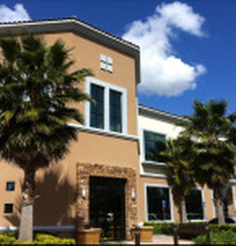
Call for an appointment
​
(951) 734-4880
Picture